
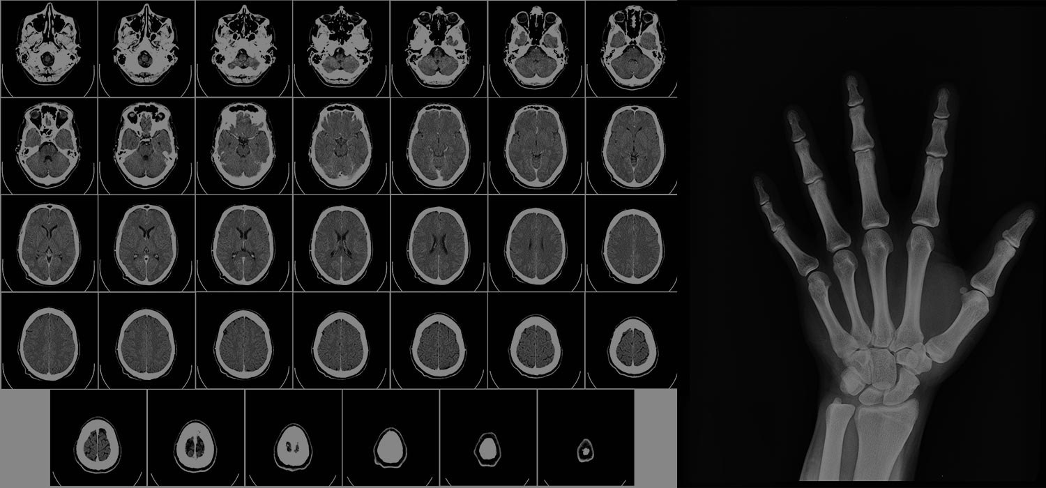
Imaging modalities, such as X-rays and CT scans, are commonly used in medical diagnosis and treatment. These imaging techniques allow doctors to see inside the body and identify various conditions, from broken bones to cancer. However, imaging modalities also have associated risks, such as radiation exposure. This blog post will discuss the benefits and risks of various imaging modalities, specifically focusing on the radiation exposure associated with X-rays and CT scans.
X-rays are a form of electromagnetic radiation that can pass through the body and create an image of internal structures. They are often used to diagnose broken bones, pneumonia, and other conditions. X-rays have several benefits, including being relatively inexpensive, widely available, and providing quick results. They also do not require an injection of contrast material and can be performed quickly.
However, X-rays also come with risks. The most significant risk is radiation exposure, which can increase cancer risk. The trouble is generally considered low, but it increases with the number of X-rays received and prolonged exposure. Children and women who are pregnant should avoid X-rays, if possible, as their developing cells are more sensitive to radiation.
CT scans (computed tomography) are another imaging modality that uses X-rays to produce detailed images of internal structures. They are often used to diagnose injuries, detect tumours, and guide biopsies. CT scans have several benefits, including providing detailed images and looking at different body layers. They also can find more minor abnormalities and injuries that a regular X-ray might miss. CT scans also can be used to detect internal bleeding, a skull fracture or other injuries in head trauma.
However, CT scans also come with risks. The most significant risk is radiation exposure, which is much higher than a standard X-ray. For example, a CT scan of the abdomen and pelvis exposes a patient to about 10 millisieverts (mSv) of radiation, while a chest X-ray exposes a patient to about 0.1 mSv. This increase in radiation exposure can increase the risk of cancer.
To minimize radiation exposure, it's important that patients only undergo imaging when it is medically necessary and that the lowest radiation dose possible is used. Also, specific protocols such as "dose modulation" can be used to lower radiation exposure. In addition, the patient needs to inform the radiologic technologist if the patient is pregnant or may be pregnant.
X-rays and CT scans are valuable imaging modalities that can help doctors diagnose and treat various conditions. However, they also come with associated risks, particularly radiation exposure. Therefore, it is essential to balance the benefits and risks of these imaging modalities and only use them when they are medically necessary. Additionally, the risk of radiation exposure can be minimized using the lowest radiation dose possible and techniques to lower the exposure.
Artificial intelligence is one such technique which can be used.
Artificial intelligence (AI) can improve imaging modalities in several ways, such as X-rays and CT scans. One of the ways it can help is by reducing the need for radiation exposure. Here are a few examples of how AI can be used in radiology to improve radiation safety:
- Automated dose optimization: AI can be used to develop algorithms that optimize radiation dose while still producing high-quality images. This can reduce unnecessary radiation exposure and lower the risk of radiation-induced cancer.
- Image analysis: AI-based algorithms can analyze images and automatically identify areas of interest, such as tumours or broken bones. This can help radiologists make more accurate diagnoses and reduce the need for additional imaging and radiation exposure.
- Computer-aided detection (CAD): AI can be used to develop CAD systems that can automatically identify and mark areas of interest in images. This can help radiologists to more easily identify abnormalities, such as small tumours, and reduce the need for additional imaging and radiation exposure.
- Quality assurance: AI can monitor image quality and identify issues that may lead to increased radiation exposure. This can help ensure that the radiation dose is as low as possible while still producing high-quality images.
- Robotic X-ray systems: AI can control robotic x-ray systems that can adjust the radiation dose, rotation, and position of the X-ray source to optimize imaging while minimizing radiation exposure.
Conclusion
It's important to note that these applications of AI are still in the early stages of development, and more research is needed to fully realize the benefits of AI in radiology fully. Additionally, these AI algorithms need to be validated, peer-reviewed and put through clinical trials to ensure accuracy, consistency and trust in the results.
